Wednesday, October 31, 2012

Cockroach: Mouth Parts

Cockroach mouth parts
  1. The remaining three pairs of cephalic appendages are small and located around the mouth. Hence together with labrum of head capsule and hypopharynx, these appendages copmrise the mouth parts of the cockroach. These help in 'biting and chewing' its food.
     
  2. Labrum: It is the broad, flattened terminal sclerite of the dorsal side of head capsule, movably articulated to the clypeus acts as upper lip. It has epipharynx (chemoreceptors) on its inner side.
     
  3. Mandibles: Thick hard and triangular appendages beneath the labrum, on each lateral side of the mouth, which bear pointed, teeth like processes called denticles.
     
  4. First maxillae: Located on each side of the mouth next to mandibles for cutting and chewing. They also bear olfactory receptors.
     
  5. Labium: The second maxillae are fused together forming a single large structure which covers the mouth from ventral side, hence called the 'lower lip' or labium. It bears tacticle and gustatory sensory setae.
     
  6. Hypopharynx: It is a small, cylindrical mouthpart, sand witched between first maxillae and covered by labrum and labium on dorsal and ventral sides respectively. It bears several sensory setae on its free end, and the opening of common salivary duct upon its basal part.

HORMONES OF ADRENAL CORTEX

HORMONES OF ADRENAL CORTEX


Adrenal glands are situated in the abdominal cavity adjacent to the kidneys. In case of fishes and amphibians the adrenal gland is found embedded in the tissues of the kidney. In reptiles, birds and mammals the Adrenal gland is distinct and independent gland located at the cephalic end of the kidney.
The adrenal gland shows two parts in various animals. The central part constitutes the medulla and the peripheral part is called the cortex. The cortex is derived from the embryonic mesoderm and the medulla is derived from the embryonic ectoderm, thus the hormones originating from them are also of a different nature.
Adrenal medullary hormones viz adrenaline (epinephrine) and non-adrenaline (non epinephrine) are of biogenic amines. The hormones are secreted under the influence of the autonomic nervous system. The Adrenaline regulates the
(i) heart beats.
(ii) Constriction-dilation of blood vessels and
(iii) Blood supply to voluntary muscles.
Adrenaline induces conversion of glycogen to glucose. It increases the rate of activity of Muscle of Trachea i.e. respiration. It retards the peristaltic movement of the wall of stomach and intestine. Adrenaline enhances production of ACTH in the anterior pituitary. Noradrenaline mainly regulates constriction/dilation of the peripheral blood vessels so as to maintain the blood pressure.

THYROID HORMONES

THYROID HORMONES:


The thyroid gland produces mainly iodine containing compounds as its hormonal secretion. Thyroxine is the most important hormone of the thyroid gland. This hormone highly influences the general metabolism of the body. The hormone has greater influence being concerned with energy transformation in the body. The thyroid hormone increases the Cytochrome-C content of the cells. Thus ATP generation is brought under its active control. This action has long-term effects on cellular metabolism.
Thyroxine is produced from Tyrosine. When the supply of iodine through diet is insufficient, the Thyroid glands swell up significantly. This leads to a disproportionate swelling of the throat region. This disease is known as “Goitre.”
Due to lack of Thyroxine
(1) Biological reactions of the body are slowed down considerably.
(2) Pulse beat becomes weak.
(3) Blood pressure is reduced.
(4) Physical as well as mental fatigue sets in quickly.
Due to excess of Thyroxine:
(1) Pulse beat increases in intensity.
(2) Blood pressure rises.
(3) The body weight decreases.
(4) Temperamentally a person becomes very irritable.

If the Thyroid gland is hypo functional right from birth then
(1) The child suffers from mental retardation.
(2) The facial features develop erratically.
(3) This leads to disfiguration.

HORMONES OF THE PITUITARY GLAND

HORMONES OF THE PITUITARY GLAND :


The hormone produced in the pituitary gland are basically proteinaceous in nature. The pituitary gland generally consists of 3 parts:

(i) Anterior Lobe
(ii) Intermediate lobe
(ii) Posterior lobe.
The hormones secreted from the anterior lobe of pituitary are described as follows.

(i) Growth Hormone (GH) or Somatropic Hormones (STH) : It regulates the overall carbohydrate metabolism and growth. Excess secretion of this hormone causes excessive growth resulting in giantism. Optimum secretion of this hormone leads to proportionate and normal growth. Very low rate of secretion of this hormone leads to dwarfism
(ii) Adrenocorticotropic Hormone or ACTH : This pituitary hormone controls the development of the adrenal cortex. This hormone influences the development and growth of the different parts(zones) of the adrenal cortex as well as medulla and regulates the production of chemicals in them.
(iii) TTH (The Thyrotrophic Hormone) or TSH (Thyroid Stimulating Hormone) controls the activity and synthesis of the hormone of the thyroid gland.
(iv) The Gonadotrophic Hormones (GTH) : This Hormone influences the growth and development of ovarian
follicles and corpus luteum. This hormone regulates the secretion of hormones by the interstitial cells of the testis. The hormone responsible for growth of ovarian follicles is known as the Follicle Stimulating Hormone (FSH).
(v) Prolactin Inhibitor Factor (PIF) : This hormone is secreted from the hypothalamus. This hormone inhibits the production of milk in the mammary glands. Thus milk is not secreted before the motherhood.
(vi) Melanocyte Stimulating Hormone(MSH) or Melanotropic Hormone (MTH) or Intermedin : This hormone is secreted from the intermediate lobe of Pituitary. This hormone is responsible for altering the skin color.
(vii) Oxytocin : This hormone is secreted from the posterior lobe of the pituitary gland. This hormone is of a polypeptide nature. Oxytocin is concerned with contraction of uterus and rapid secretion of milk from the lactating, mammary gland of women.
(viii) Vasopressin or Antiduretic Hormone (ADH) : This hormone is also secreted from the posterior lobe of the pituitary and is of polypeptide nature. This hormone controls
(i) viscosity of arterial blood.
(ii) the constriction of arteries and
(iii) osmoregulation in kidney and body.

Animal Hormones

Animal Hormones:

Hormones are produced in the endocrine glands of animals. The pituitary gland and hypothalamus are the most important in regards to control and development. The pituitary gland is responsible for the production of a hormone called somatotrophin. Somatotrophin is essential in the fact that it promotes mass production of proteins on a body-wide scale, by accelerating the rate of transport of amino acids; the constituents of a protein.
The same part of the pituitary gland is responsible for thyroid stimulating hormone or TSH for short. This targets the thyroid gland, also a member of the endocrine system, which in turn promotes the production of thyroxine. Thyroxine is responsible for controlling the body's metabolic rate, and therefore responsible for the amount of energy consumed and the volume of proteins produced.

PROPERTIES OF HORMONES:


Hormones are responsible to a great extent for either acceleration or retardation of enzyme action and its production. In this manner hormones regulate physiological activities through chemical co-ordination. Certain secretions 'Hormones' of this endocrine glands are conducted through the circulating blood. The glands which produce such secretions do not possess ducts, hence the endocrine glands are also called ductless glands. A hormone belonging to the individual of one species may be similar to the corresponding hormone of the individual of other species. This is a special characteristic of hormones.
CLASSIFICATION OF HORMONES

Hormones are classified into various types on the basis of their chemical structure as under:
(1)  Steroid Hormones: Estrogen, Progesterone, Aldesterone, Cortisone etc.

(2)  Biogenic Amines Hormones: Adrenaline, Noradenaline, Acetylchloline etc.

(3)  Peptide/Protein Hormones:Thyroxine, Glucagon, Insulin, Thyrotropin, Gonadotropins,
     Adrenocorticotropin,
     Parathormone etc.
Hormones are secreted in very small amounts. Hormones are chemicals which help in the regulation of various physiological activities like metabolism, growth, reproduction, metamorphoris, ecdysis or moulting, formation of pigment granules and osmoregulation in the body. The regulatory functions of the endocrine system are observed to be controlled either directly or indirectly. In animals it has been observed that the central nervous system influence the hormones regulating the color changes of the skin.

The secretion of gastric hormones are stimulated:
(i)  Under the influence of the nervous system and the (ii) local biochemical variations in the organs.
These gastric hormones regulate the secretion of the digestive enzymes.

Frog: Copulation

COPULATION IN FROGS

 http://images.joelsartore.com/preview/A/ANI025-00218.jpg
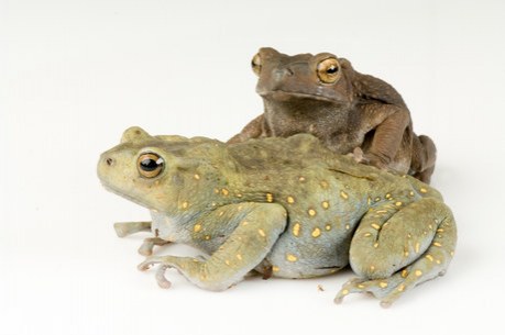
The entry of the sperm in the egg is the basic requirement of sexual reproduction. The eggs are enveloped by liquid viscous albumin. When this albumin comes in contact water, it swells by absorbing it (water). This Albumin becomes very thick and transparent, colorless jelly. This jelly does not permit the entry of any sperm through it. The fertilization is external and in water. So it essential that the sperm should be able to enter the egg before the jelly formation. For this purpose the sperms should remain in as close a contact with egg is possible. The copulation is essential in frogs, in order to provide the maximum possible number of eggs, an opportunity for fertilization within a very short time. The frogs reproduce in monsoon in dim light. Usually when it is raining the male frogs collect near the bank of water pool or ditch. They produce croaking sound with the help of vocal sacs. The vocal sacs act as amplifiers. The croaking is a mating call for the female frogs. The female frogs getting attracted approach the male frogs. The male frog rides over the female frog and embraces it. The male frog holds the female frog firmly by its forelimbs and nuptial pads. The couple remains in this condition for 2-3 days. The Frog takes a long time to become sexually excited, as they are cold-blooded animals and devoid of copulatory organs. The male holds the female more tightly at the state of orgasm. At this stage the female discharges a large number of eggs in water from its ovisac through the cloacal aperture. The male frog right at the same moment discharges its sperms over the eggs falling in water. The two animals separate from each other on completion of this process.

Frog: Fertilisation

FERTILIZATION IN FROGS


The process of copulation is immediately followed by the process of fertilization. The fertilization is external and in water. Each egg falling in water has a small conical protuberance from the equitorial plane. This conical protuberance is called reception cone. The tip of the reception cone is thin walled and projecting out of the albuminous layer. Normally, each egg is surrounded by innumerable sperms. The first sperm that reaches the tip of the reception cone dissolves the tip with the help of an enzyme secreted by the acrosome on its head and makes its way in to the egg. The head and middle piece of the sperm enter the egg, while the vibratile tail is cut off and remains outside the egg.
During fertilization a series of changes occur in the egg. The nucleus of the secondary oocyte divides into two nuclei.
(A)One of these nuclei remains in the egg cell. It is known as female pronucleus. The other nuclei is pushed out through the egg cell membrane. This nucleus settles as second polar body. It remains near the first polar body.
(B)The reception cone becomes flattened and closed. The cell content of the egg with egg cell membrane shrinks. The cell content of the egg gets fully separated from the egg membrane. The egg membrane is now termed as fertilization membrane.
(C)The head of the sperm that has entered the egg changes its shape. The head of the sperm becomes a round male pronucleus.
(D)The male pronucleus moves towards the female pronucleus and finally fuses with it to form a dioploid (2n) nucleus called zygote nucleus. The melanin granules settle deeper from the surface exactly on the opposite side of the site of entry of the sperm in the egg. As a consequence a much less dark (grey) colored semilunar patch called grey crescent is formed. The egg having grey crescent formed in it is recognized as a fertilized egg or zygote.

IMPORTANCE OF FERTILIZATION


Generally the unfertilized egg does not develop into embryo and produce new animal. Fertilization is thus essential. In the process of fertilization the haploid egg and the haploid sperm produced during gametogenesis meet and fuse with each other. This establishes the diploid state in the zygote. Thus, the specific diploid number of chromosomes of the species is maintained. The maturation of the egg cell is completed only after the entry of the sperm in it. The process of fertilization adds new vigor and activeness in the egg cell. The egg cell becomes active for embryo development through the process of fertilization. The process of fertilization brings about the combination of characters of both the parents in the off springs. The grey crescent formed in the process of fertilization determines the future anterioposterior axis and the axis of bilateral symmetry in the spherical zygote.

Frog: T.S. Testis

MICROSCOPIC STRUCTURE OF TESTIS


 Testis(internal-structure).JPG
Histological the testis consists of numerous
1. Seminiferous tubules
2. Blood vessels
3. Nerve fibers and
4. Interstitial cells embedded in connective tissue.
All the mentioned above are enclosed by a common, thick fibrous sheath of connective tissue. This sheath is called Tunica Albugenia. The seminiferous tubules are lined by a single layer of cuboidal cells. These cuboidal cells form the germinal epithelium. The germinal epithelium produces a large number of sperms through the process of spermatogenesis. The sperms can be seen in the form of several bundles. These bundles are radially arranged in the lumen of each seminiferous tubule. Some cells in the process of spermatogenesis are also seen in the lumen of seminiferous tubules. The connective tissue lying in the intertubular spaces contains one or more interstitial cells along with blood vessels and nerve fibres.The interstitial cells are also called Leydig's cell. The interstitial cells are endocrinal in nature. They secrete male sex hormones, such as androsterone and testosterone.

Frog: T.S. Ovary

MICROSCOPIC STRUCTURE OF OVARY


 Ovary(Internal-structure).JPG

Histologically, the wall of the ovary is made up of
1. An outer epithelium,
2. An inner epthelium,
3. Connective tissue,
4. Blood vessels and
5. Nerve fibers.
The outer epithelium and the inner epithelium are connected with each other by connective tissue. The outer epithelium is formed of flattened cells. At certain intervals in these flattened cells there are somewhat larger cells that participate in the formation of ovarian follicles. These larger cells form the germinal epithelium. The germinal epithelial cells form ovarian follicles through the process of cell multiplication and growth. These ovarian follicles are of different sizes. This difference in size depends upon the level of their development. Each ovarian follicle is formed of a large and centrally located developing egg cell (primary oocyte). The egg cell (primary oocyte) is surrounded by follicular cells and almost completely enclosed in the inner epithelial layer of the ovary. The inner epithelium is uniformly thick and made up of flattened epithelial cells.

Reproductive system of frogs

Reproductive system of frogs

The male frog and the female frog can be distinguished even by their external morphological characters. The organs concerned with the production of gametes [sperms and ova (egg)] are called principal reproductive organs. The organs concerned with the transport of gametes further from the gonads are called accessory reproductive organs.

REPRODUCTIVE ORGANS OF MALE FROG

In the male frog the principal reproductive organs are a pair of testes and the accessory reproductive
organs are :
1. Vasa Efferentia,
2. Bidder's Canal,
3. Collecting Tubules,
4. Urinogenital Ducts,
5. Cloaca and
6. Cloacal Aperture.
 Urinogential-system-of-male-frog.JPG

  1. Male reproductive organs consists of a pair of yellowish ovoid testes, which are found adhered to the upper part of kidneys by a double fold of peritoneum called mesorchium. Vasa efferentia are 10-12 in number and after arising from testes run through the mesorchium and enter the kidneys of their side. In kidneys, these open into Bidder's canal, which finally communicates with the urinogenital duct.

    This duct emerges from the kidneys and finally opens into the cloaca. The cloaca is a small, median chamber that is used to pass faecal matter, urine and sperms to the exterior. 
 
REPRODUCTIVE ORGANS OF FEMALE FROG
 

In the female frog the principal reproductive organs are a pair of ovaries and the accessory reproductive organs are :
1. A pair of Oviducts,
2. Cloaca and
3. Cloacal Aperture.
Ovary : In the young frog each ovary is small, flat and lobulated. In adult frog this is the same case during the period of year other than breeding season. The ovary is enveloped by mesovarium, a thin layer of peritoneum. Several hollow sac-like structures form the ovary. The lobulated appearance of the ovary due to those structures. Initially the colour of the ovary is yellowish with small black spots. The lumen of the ovary is part of the coelom. It is filled with coelomic fluid. During the breeding season the wall of the ovary becomes studded with a large number of ovarian follicles. Each Ovarian Follicle contains a developing Egg. The ovarian follicles project towards the lumen of ovary. Such an ovary greatly enlarges. It attains black color with light yellow spots. Each oviduct is a long narrow and highly coiled tube. It is divided into three parts in accordance with its structure and functions.
(1) Oviducal Funnel,
(2) Oviduct and
(3) Ovisac.
The anterior end of the oviduct forms a wide and fringed oviducal funnel. The ovoiducal funnel is located on the dorsal side of the lung. The margin and inner surface of the oviducal funnel is lined by ciliated epithelium. The oviducal funnel leads into the oviduct. This oviduct is straight and thin-walled for a short distance. Thereafter it becomes highly coiled and thick-walled. This coiled oviduct runs posteriorly along the outer side of the kidney. The hinder portion of the oviduct becomes very thin walled. It is sac-like and is called ovisac. The ovisac opens of the posterior end in the dorsal wall of the cloaca by its individual apertures lying anteriorly to the openings of ureters. The cloaca opens to the exterior by a cloacal aperture at the posterior end of the body.
The release of ovum in female is termed as spawning.

Respiratory System of Frogs

Respiratory System of Frogs
  1. Three types of respiration: cutaneous, buccopharyngeal and pulmonary.
  2. Cutaneous respiration (35% of respiration) is through the body surface. During the hibernation and aestivation, frog respires only through this method.
     
  3. Buccopharyngeal respiration occurs through the lining of buccal cavity. It occurs only when frog is out of water (0.9% of total respiration). The mucus mebrane of the buccal cavity is moist which dissolves oxygen and diffuses it into the blood capillaries.
  4. Pulmonary respiration: Lungs in frogs are not efficient respiratory organs because only mixed air enters into them and mainly function as hydrostatic organs. Lungs are pair of thin walled, translucent with inner surface divided into alveoli by septa. Pulmonary respiration has a maximum frequency of 20/minute. It occurs when more energy is required. Mouth and gullet are kept closed during pulmonary respiration.


    Respiratory movements in pulmonary respiration are because of buccopharyngeal cavity which acts as a force pump. These movements are carried out by set of paired muscles - sternohyal and pterohyal muscles. Sternohyal muscles are attached with hyoid and coracoid, clavicles of the pectoral girdle and on contraction depresses the buccal floor enlarging the buccopharyngeal cavity. Pterohyals are attached in between hyoid and pro-otics of the skull and on contraction lifts the floor of buccal cavity. With the depression of buccal floor, air enters buccal cavity through the nares. External nares are then closed by pushing tuberculum prelinguale and the movable premaxillae. It is followed by raising of the buccal floor by pterohyal muscles which reduses the volume and air is pushed into the lungs where exchange of gases takes places. Buccal floor is again lowered enlarging its volume which draws air into the buccal cavity. External nares are opened followed by raising the buccal floor, pushing the air out through external nares.
     
  5. Sound producing organs of frog is laryngo-tracheal chamber. It is supported by one cricoid, two arytenoids and two prearytenoids. It has a pair of muscle strands vocal cords which actually produse sound. Male frogs has vocal sacs which act as resonating chambers.

Animal Husbandry

Animal husbandry deals with the breeding, feeding and caring of domestic animals. Like any other farming practice; the production and management of live stock, requires definite planning for their shelter, nutrition, breeding, disease control, and proper economic utilization. Among milk producing milch breeds of cattle; Red Sindhi, Sahiwal and Gir are the main indigenous varieties while Jersey, Holstein-Friesian and Brown Swiss are very important exotic breeds of dual purposes. National Dairy Research Institute Karnal, Haryana develops Karan Swiss, Karan Fries and Frieswal cross breeds of very high milk producing capabilities. Among buffaloes Murrah, Mehsana and Surti are high milk producing variety produces 1800-2500 litres 1200-2500 litre and 1600 to 1800 litre milk respectively. Proper shelter and food is required for dairy animals to manage their good health and dietry requirements.

FMD, Anthrex, rinderpest, haemorrhagic septicemia, black quarter and cow pox are some common disease of domestic animals. Effective vaccines against all these major viral and bacterial disease have been developed and are available in our country, Poultry farming, Fish farming and meat providing livestocks are another new fields of animal husbandry.

In India the popular indigenous breed of poultry includes Aseel or (Indian game), Peela (golden red), Yakub (Black and red), Nurie (white) and Kajal (black) etc. and white leghom, Rhode island red are the common exotic breeds which are good egg layer and also meat yielding (broiler).

Fish is another important proteinaceous food resources. There is good scope of producing fish in our country. Our inland fishery includes 1.6 million hectares and our long coast line of 6,500 km provides a high potentials for fish production. Catla, Rohu, Mrigal with some exotic varieties like silver carp and grass carp are important edible fishes. Some disease like IPN (Infectious Pancreatic Necrosis) and VHS (Viral Haemorrhagic Septicemia) are well known infectious disease of the fish. Water pollution causes great harm to fish.

Besides poultry and fishes; goat, sheep and pig combined together constitute about 70% of the total meat supply in India. Nellore and Mandya are famous for production of wool, Dorset and Suffolk are popular cross-breed variety. About 20 variety of goat are found in our country; Jamunapari, Himalayan, Bengal and Assam hill breed are popular indigenous breed while Alpine, Toggenberg and Sannen are exotic breeds. disease like black quarter, Brucellosis and vibriosis are common bacterial disease of goat and sheep. Sore mouth, goat pox and rinderpest are their common viral disease.

Human Diseases

According to World Health Organization (WHO) health is defined as a stage of complete physical, mental and social well being and not merely the absence of disease or infirmity and the term disease means dis ease or without ease or discomfort. Therefore the disease ca be defined as the malfunctioning of the body organs due to one reason or the other. The human disease can be broadly categoriesed in Communicable (infectious) and non-communicable (Non infectious) disease.

Among the communicable disease, malaria is caused by Plasmodium. Headache, nausea, muscular pains and high fever are its main symptoms. To protect from the disease one should have to protect himself from the mosquito bite and use of antimalarial medicines like Quinine, Paludrine. Metakalfin etc.

Influenza (Flu) is highly infectious disease caused by Myxovirus influenzae. Sneezing, coughing, fever, are its main symptoms. There is no effective control for influenza is available. Jaundice or hepatitis spread mostly through contaminated food and water with hepatitis virus. High fever, headache, loss of appetite with dark yellow urine are its main symptoms. Rabies (Hydrophobia) spread through bite of a rabies dog, Fox, Cats mainly. Severe headache high fever, and painful contraction of muscles of throat and chest. Severe damage to CNS causes paralysis and painful death. AIDS is caused by HIV virus. It causes secondary immune deficiency disorders. It is preventable but not curable. Loss of weight diarrhea, fall in Lymphocyte count are the main symptom.

Tuberculosis s caused by Mycobacterium tuberculosis. Here patient feels sick and weak. Loss of weight, chest pain and breathlessness are common features. Use of BCG vaccine protect from disease. Cholera is common during fairs and after flood. It is caused by vibrio cholerae and spread by contamination, Immunization of crowded place, use of boiled water personal hygiene helps in prevention. Use of ORS and anti bacterial medicines is suggested.

Typhoid is the most common communicable disease of India; occurring in the age group of 1-15 years mainly. It's caused by salmonella typhi. Headache and high fever, continues. TAB vaccination, provides immunity for 3 years. Typhoral vaccine also prevents typhoid. Diarrhea is mainly caused by some type of bacteria, protozoa and virus. Frequent loose motions and vomiting, leading to dehydration. Use of ORS, husk of isabgol seed, pulp of unripe banana and antimicrobial drugs provide relief.

Among non-communicable disease Marasmus, Kwashiorkor is mainly caused by protein deficiency while anemia and goitre is caused by deficiency of Iron and Iodine respectively.

Xeropthalmia, Beri-Beri, Rickets, Pellagra, Scurvey are the important vitamin deficiency disease. These disease can be cured by proper dietry supply.

Reproduction

Reproduction

Reproduction is creation of new individuals belonging to the same species. Though reproduction is not necessary to maintain life of an individual but maintains the species or population . Reproduction can be said to involve making copies of blue prints of body design. The basic event in the reproduction is the creation of DNA copy.

For cellular organisms, cell division or fission, leads to formation of new individuals. Fission can be binary if forming only two daughter individuals e.g., Amoeba, Paramecium otherwise if divide into many daughter cells simultaneously, it is called multiple fission e.g., Plasmodium. Binary fission can occur in any plane e.g., Amoeba or in a definite orientation in relation to their structures.

This is not true for multicellular organisms which need to use more complex ways of reproduction. Some organisms like Hydra can use regenerative cells for reproduction in the process of budding. A bud develops as a small outgrowth and can develop into small individual which when fully mature, get separated from the parent body and become new individual. This type of reproduction is called asexual reproduction and a single parent is involved.

Other mode of reproduction is called sexual reproduction. It involves participation of both, male and female sexes. Human use a sexual mode of reproduction only.

Human Reproductive System
1.Male reproductive system: It consists of 2 parts
  1. Portion which produce the germ cells/spermatozoa (Testes)
  2. Portion which deliver spermatozoa to the site of fertilization.
A pair of testes are located in the scrotal sac, outside the abdominal cavity. This is to provide optimum temperature for the formation of spermatozoa which is slightly lower than the body temperature. Besides, testes are also an endocrine structure releasing testosterone. Testosterone is involved in appearance of secondary sexual characters and also, regulates formation of sperms.

The sperms from the testes pass through vas deferens. Both vasa deferentia combine with each other and also with a tube coming from the urinary bladder to form urethra. Thus, urethra is a common passage for sperms and urine. Along the path of vas deference secretion from seminal vesicle and prostate gland gets added forming the semen. As the sperms are now in a fluid, this makes their transport easier. The semen also contains fructose to provide energy. The sperms are the flagellated structure having a long tail. This helps them to move towards germ cell or ovum.
2.Female reproductive system: Female reproductive system has a pair of ovary for producing germ cells/eggs/ova. Besides, ovary also produce oestrogen hormone responsible for development of secondary sexual characters at puberty. At the time of birth, ovaries already contain thousands of immature eggs. Maturation of some eggs starts phase-wise on attaining puberty. One egg is produced every month by one of the ovaries. From the ovary, ovulation or release of ovum occurs which enters the thin oviduct/fallopian tube. Both the oviduct open into womb/uterus, which is an elastic bag like structure. The uterus open through the cervix into the vagina.
3.Fertilization and development: During sexual intercourse, the spermatozoa are released into vagina and from these, these move upward and reach the fallopian tube. The sperms encounter ova in the fallopian tube and fertilization occurs forming the zygote. Zygote starts dividing (cleavage) and also move towards uterus to get implanted in the lining of uterus. Rest of development of embryo/foetus occurs in uterus. The embryo gets nourishment from the maternal blood with the help of a special tissue called placenta. The waste generated by developing embryo also gets removed through placenta into mothers blood. The development of child inside the mother's body takes approximately nine months (Gestation period). The child is born as a result of rhythmic contraction of the muscles in the uterus.

If the eggs is not fertilized, it survives for about 24 hours. As one ovum is released each month by ovary, the uterus also prepared itself each month to receive a fertilized egg. Uterus gets prepared by making its lining thick, spongy and highly vascular. If implantation fails, the thick lining of uterus is also lost and comes out through the vagina as blood and cells. This is called menstruation. It usually lasts for 2-8 days and also takes place roughly every month.
Reproductive Health
As sexual act is very intimate contact between the bodies, a number of disease can get transmitted sexually like gonorrhoea, syphilis, warts, HIV-AIDS (sexually transmitted disease). The transmission can be prevented to certain extent by using condom which is a covering for penis.

Contraceptive methods: Many ways have been devised to avoid pregnancy during sexual contact. These can be
  1. Mechanical barrier: These prevent the contact between sperm and ova. Condoms on the penis or female condom worn in vagina.
  2. Hormonal methods: By changing the hormonal balance which prevents release of egg fertilization. These can have side effects also.
  3. Intra-uterine devices (IUD): These are placed in the uterus e.g., Loop or Copper T.
  4. Surgical approach: By blocking the vas deferens in male or fallopian tube in female. This method is safe in the long run.
Surgical methods can be used for removal of unwanted pregnancies (Medical Termination of Pregnancy-MTP). This may be misused for female foeticide. Because of reckless female foeticides, child sex ratio is declining at an alarming rate. For that reason prenatal sex determination has been prohibited by law. For a healthy society the female-male sex ratio must be maintained.

Evolution

Evolution

1.The diversity of living world is enormous and a large number of them have not been identified so far. Not only this, we also know about the fossils, which are the remains of dead organisms, which lived in remote past. How and from where did such a wide variety of organisms come to exist? How did human evolve? The branch of biology that deals with all these problems is called evolution.

Evolution is a slow, continuous and irreversible process.

Origin of life:
J.B.S. Haldane, a British scientist (who became a citizen of India later), suggested that the first life developed from the simple inorganic molecules which existed on primordial earth. The primordial earth had a reducing atmosphere and was rich in CH4, NH3 and H2 and water. Harold C. Urey an astronomer asked his student Stanley L. Miller to perform an experiment to prove Oparin and Haldane's hypothesis. He constructed an apparatus of glass tubes and chambers. In the spark chamber he added CH4 : NH3 : H2 in the ratio of 2 : 1 : 2. After 18 days, significant amount of the simple organic compounds such as amino acids appeared. Amino acids formed were alanine, glycine and aspartic acid.
2.Evidences of Evolution
Homologous organs are those which have the same basic structure and developmental origin but perform different functions. The organisms which possess such organs are said to have originated from common ancestor.

Example: The forelimb of a frog, a bird and a human being, show structural similarity; but the function of these fore limbs is entirely different from that of each other. The similarities indicates that all these vertebrates had common ancestor.

The wing of insects and birds are structurally different but perform the same function i.e., flying. They are termed as analogous organs.
3.If we study the embryos of different groups of vertebrates, they show striking similarities. All have gill slits, notochord and tail. This laid Ernst, Haeckel to propose a law, called as biogenetic law. The law, states that "Ontogeny recapitulates phylogeny. Which means that during the embryonic development of any organism, it repeats ancestral history".
4.Vestigial organs are functionless remnants of the organs which were once functional in ancestors e.g., vermiform appendix, nictitating membrane of human eye. Appendix is remnant of large caecum, functional in reptiles.
5.Fossils also provide evidence of evolution for example, the fossil Archaeopteryx looks like a bird, but it bears a number of other features, which are found in the reptiles. This indicates birds have evolved from reptiles.
Theories of Evolution
6.Jean Baptiste Lamarck, gave the theory of inheritance of acquired characters.

The theory was put forward in the book Philosophies zoologique.

According to this theory use of an organ leads to strengthening and disuse leads to weakening. All these changes or variations are inherited by the offspring's. August Weismann, discarded the theory of inheritance of acquired characters.
7.Later, Charles Robert Darwin, had put forward the theory of natural selection or Darwinism, in his famous book "The Origin of Species". "Darwin was most influenced by the essay on population" written by Malthus. He made the observations of nature during his voyage on the ship H.M.S Beagle. According to Darwin all the organisms have a high reproductive potential, but there is constancy in number. There is struggle for existence in nature and due to variations there is survival of fittest.
8.Now the most accepted theory of evolution, is known as synthetic theory of evolution, in which origin of spices is based on the interaction of genetic variation i.e., population genetics and natural selection.
9.Natural selection is not the only force responsible to bring about changes in gene frequencies. There is the role of chance or genetic drift also e.g., in the environment on bushes beetles are present. They show variations, most of the beetles are green in color, some red and some blue, Crows can not see green colored beetles on the green leaves of the bushes, and therefore can not eat them. This variations becomes common because it had survival advantage and thus was naturally selected. Natural selection is exerted by crows. Suppose an elephant comes and stamps on the bushes where beetles lives. This kills most of the beetles by chance, the few beetles that have survived are mostly blue. Now blue colored beetles become common, through there is no survival advantage. So genetic drift, provides diversity without any adaptations.
10.Micro-evolution occurs at genetic level due to differences in the alleles of the same gene. They simply change the common characteristics of the species.
11.Speciation may take place when variation is combined with geographical isolation. Example, if between the two sub-populations of beetles a large river comes into existence the two populations are further isolated. The levels of gene flow between them will decrease. Over generations, genetic drift will accumulate different changes in each sub-population.

Together, the process of genetic drift and natural selection will result in these, two isolated sub-populations of beetles to become more and more different and slowly give rise to new spices.
12.Artificial selection some genetic variability is always present in the population, Some alleles make organisms live and reproduce successfully. In artificial selection the role of nature is played by man, e.g., cabbage, cauliflower, kohlrabi are descendants of a common ancestor colewort, obtained through artificial selection.
13.Human Evolution: Study of human evolution indicates that all of us belong to a single spices that evolved in Africa and spread across the world. The earliest members of the human species, Homo sapiens, have been traced in Africa. So, all human races have originated from a common Homo sapien ancestor in Africa.

Glands:

Glands:


Glandular epithelium are specialized for secretory activity.
A cell, tissue or organ which secretes a useful chemical material is known as gland.
Glands are made up of cuboidal epithelial cells which are more secretory.
All glands arise as folding of epithelia. The golgi body in gland cells are larger and more secretory.
Most of the glands of body are merocrine types. It originate from all three germinal layers. (ecto, meso and endoderm).  
Liver is the largest gland of the body and lined by glandular epithelium.

Types of glands:

    • Unicellular gland : It  consist of unicellular gland cells which are called as goblet cells or chalice cells. They secrete mucous and found in mucosa of intestine and stomach. Mucous lubricates the food for easy peristalsis. Their life span is about 2–3 days.
    • Multicellular gland : It consist of many cells and are generally located in underlying connective tissue e.g. gastric and intestinal glands.
    • Exocrine gland : These are those glands which discharge their secretory products into ducts. It is also called ducted glands or glands of external secretion.  e.g. Salivary glands, Mammary glands and Tear glands.
    • Endocrine gland : It is often called ductless gland, because they discharge their secretory products (hormones) directly into the blood. e.g. Pituitary gland, thyroid, parathyroid and adrenal glands.
    • Heterocrine gland : These are those glands which are partly endocrine and partly exocrine in function. e.g. Pancreas.
    • Structural classification of exocrine glands : Multicellular exocrine glands are classified by structure, using the shape of their ducts and the complexity (branching) of their ducts system as distinguishing characteristics. Shape include tubular and alveolar (Sac like). Simple exocrine glands e.g. intestinal glands, mammalian sweat glands, cutaneous glands of frog etc. have only one duct leading to surface. Compound  exocrine glands have two or more ducts e.g. liver, salivary glands etc.
    • On the basis of mode of secretion, glands can be:
       
    • Holocrine glands: In holocrine glands (e.g., sebaceous gland), the product of secretion is shed with the whole cell leading to its destruction.
       
    • Merocrine glands: When the secretory granules leave the cell by exocytosis with no loss of other cellular material, the glands are called merocine glands (e.g., the Pancreas).
       
    • Apocrine glands: In aporcrine glands (e.g., mammary gland and axillary sweat glands), only the apical portion of the cytoplasm is discharged along with the secretory product.

    •  

Epithelial Tissue:

Epithelial Tissue:


  • An epithelium is a tissue composed of one or more layers of cells that cover the body surface and lines its various cavities. 
  • It serves for protection, secretion and excretion. 
  • The word ‘epithelium’ was introduced by Ruysch
  • It was applied originally to thin skin covering the nipple (G. epi = upon, thele = nipple). 
  • They are located on the outer surfaces of organs, including the skin. 
  • They form the linings of tracts, cavities and vessels.  
  • Epithelial tissue evolved first in animal kingdom. It originate from all the three primary germ layers. e.g. Epidermis arises from ectoderm, Coelomic epithelium from the mesoderm and epithelial lining of alimentary canal from the endoderm.


Structure:
  • Cells are arranged in one or more layers, cells are compactly arranged and there is no inter cellular matrix between them. 
  • Neighbouring cells are held together by intercellular junctional complexes like desmosomes, tight junctions, interdigitations etc. the cells of lowermost  layers always rest on a non living basement membrane or basal lamina.  
  • Basement membrane is made up of no cell product of epithelial tissue. 
  • It is formed of mucopolysacharides, glycoprotein and collagen or reticular fibres. 
  • The epithelial cells are held together by small amounts of cementing substances is mainly composed of glycoprotein secreted by the cell themselves. 
  • Blood vessels are absent in the epithelial tissues. 
  • However, nerve endings may penetrate the epithelium. 
  • The free surface of cells may be smooth or may have fine hair like cilia, sterocilia and microvilli. 
  • Epithelium is subjected to continuos wear and tear and injury. Hence it posses very high capacity of renewal (mitotic cell division). 
  • The following types of modifications and junctions are found in the plasma membrane of adjacent epithelial cells to keep the cells together.
 Modifications in epithelial tissue
  • Microvilli : It is simple and minute cytoplasmic processes arising from free exposed surfaces of the cell. They absorb material.
  • Stereocilia : It is non-motile cytoplasmic processes.
  • Cilia : It is contractile motile fibrous processes arising from basal granules.
  • Tight junctions  (Zona occludens) : At certain places the plasma membranes of adjacent cells are tightly packed or even fused together.
  • Desmosomes : Desmosome is present in epithelial tissue. They consist of thickened area and several fine tonofibrils extending from  each plasma membrane into cytoplasm of respective cells. Macula adherens is a kind of desmosome.
  • Gap junction : At place, the adjacent cells form ion-rich gap junctions for intercellular communication and chemical exchange. These junctions probably do not provide physical support.
  • Interdigitations : These are interwoven finger-like processes of plasma membranes of adjacent cells.
  • Intercellular bridges : These are minute projections that arise from adjacent cell membranes. The intercellular bridges make contact with one another.

Tissues

Tissues


  • The word “tissue” was given by French anatomist and physiologist Bichat (1771-1802).
  • “Tissue” was earlier used by N.Grew (1682) in connection with plant anatomy.
  • Study of tissues is called histology. (Gr. histos-tissue, logos-study).
  • Term ‘histology’ was given by German histologist Mayer (1819).
  • Italian scientist Marcello Malpighi (1628-1694) is known as “founder of Histology”
  • Histology is also called as microscopic anatomy (microanatomy).
  • Bichat (1771-1802) is considered “Father of histology”.
  • Histogenesis (histos-tissue, genesis-formation). Formation and development of tissues from undifferentiated or embryonic cells also called histogeny.
  • The phenomenon of differentiation of cell groups into tissues is known as histodifferentiation.
  • Histolysis (GK. histos-tissue, lysis-dissolve) Degeneration of tissues.

Ageing

Ageing


(i) Definition : Ageing is the show deterioration in the structure and function of body cells tissues and organs of an animal and starts after the adulthood.
(ii) Gerontology : The field of developmental biology that deal with the process and problems of ageing is known as gerontology – (Gr. geron = old man; logos = discourse). The scientists involved in the science of ageing are called gerontologist.
(iii) Life cycle and life span : In all metazoan animals, the life cycle includes two developmental period; embryonic period (pre-natal developmental period) which extends from zygote to offspring till hatching or birth, and post embryonic period (post-natal developmental period)- which includes growth, adulthood, reproduction, ageing. Thus, the life cycle comprises five main events : birth, growth, maturity, old age and death, that follow in the sequence named. Maximum life span is the maximum number of years survived by any member of a species, while average life span is the number of years survived by members of a population. Life expectancy is the age at which half the population still survives. The life span varies greatly in different organisms :
Maximum life span of human has been found to be about 121 years. Shirechiyo Izumi of Japan died due to pneumonia at the age of 120 years and 237 days in 1986.
Average life span of women is longer than men while the biological process of ageing is faster in human male than in human female. No organism lives for ever. (It is believed that Hydra is immortal as it does not undergo ageing). Every organism dies of old age if not killed earlier by an accident, a predator, aparasite or a disease.
(iv) Why old age ends in a natural death : Though it is difficult to give a categorical answer, certain factors that lead to death in old age are known. During the growth period, new cells are formed faster than the rate of death of old cells. But after the maximum growth, the metabolic rate declines and rate of formation of new cells is lower than the rate of death of body cells. So the repair of damaged cells is not complete and a slow deteriorative process starts. In human beings, it starts after the age of 30 years. But in certain cases, deterioration in structure and function of cells may start during childhood or even during prenatal life e.g. hearing efficiency of ear and atresia in ovary.  As the organism grows older,
(a) The metabolic activities gradually decline.
(b) Capacity to replace the worn out cells decreases.
(c) The power to repair the worn out tissues and organs decreases.
(d) Resistance power to diseases is lowered.
(e) Lowered adaptability.
(f) Finally, non-functioning of some vital organs like heart, brain, kidneys, lungs or liver.
So with ageing, there is an impairment of physiological functions, called senescence and ultimately leads to death.
Peak of activity : In humans, generally speaking, the peak of activity is thought to reach at about 30 years of age. Thereafter, the body shows signs of senescence, i.e., deterioration and loss of normal function. From the age of about 30 years, the human body becomes functionally less efficient by about 0.8% every year. It is a biological process.
(v) Deterioration data of an old person : A 75 years old man, for instance, has, as compared to a 30 years old person, about 64% less taste buds, about 44% less renal glomeruli about 20% less nerve cells in the brain, and about 37% less axons in the spinal nerves. His heart pumps 35% less blood and sends 20% and 58% less blood to the brain and the kidneys respectively. His lungs have 44% less vital capacity and provide about one-third less oxygen to the blood per minute. His kidneys have 31% lower rate of glomerular filtration. His nerve impulses are propagated at a rate about 10% slower. At the age of 30, the height starts decreasing indetectibly, it decreases by 0.3 cm. at 40, by 2 cm. at 50 and by 2.5 cm. at 70.
(vi) Changes in ageing or symptoms of ageing : Gerontalogist have worked out a large number of changes that accompany ageing. These are discussed below under three heads –
(a) Changes at organ level : During ageing, different organs and organ systems show different rates of decline e.g.
(1) Heart : With increasing age the efficiency of heart decreases. In a man of 70 years, the heart pumps only 65 per cent blood per minute as compared to a 30 years old man. Consequently, the blood going to the brain and kidney is reduced to 80 percent and 40 percent respectively.
(2) Oxygen uptake by blood : At the age of 20 blood takes about 4 litres of oxygen per minute, while in a man of 75 years, it takes only about 1.5 litres of oxygen in the same period.
(3) Decrease of blood volume : The production of new RBCs from the bone marrow declines and consequently the volume of blood also decreases.
(4) Kidney : The number of kidney tubules is found to reduce to half in the old age. As a result the volume of urine decreases. This creates lots of other urinary troubles and also causes body ache, low back and difficulty in passing urine.
(5) Lungs : The capacity of lungs for intake of air decreases. This leads to reduction in the oxygen supply to different tissues. Therefore, old persons suffer from breathlessness and inflammation of mucous membrane.
(6) Digestive system : The number of taste buds on tongue reduces to about one-third. The secretion of digestive juices also decreases with old age. This may result in indigestion. loss of appetite, dyspepsia, constipation and gas formation.
(7) Retention of water : The capacity of body cells to retain water also decreases with the result, the skin in old persons is dry and wrinkled.
(8) Nerve impulse : The rate of nerve impulse propagation reduces with age. The decline is about ten percent in man of 75 years as compared to that of 50 years old person.
(9) Increased mineral deposition in the bones which become brittle and easily fracturable.
(10) Muscle tissue degenerates due to biochemical changes in the muscle cells or neuromuscular junction. This greatly reduces the muscular strength. Without exercise, estimated muscle mass declines 22 percent for women, and 23 percent for men. It can be prevented by regular exercise.
(11) Thymus is almost microscopic by the age of 70 years. This lowers the number and functioning of T-lymphocytes which lowers the immunity against the antigens.
(12) Hair start greying or falling at the age of 40 due to reduced rate of protein synthesis.
(13) Menopause in female at an average age of 52 years.
(14) Body becomes thin, shrivelled and stooping.
(15) Decline in hearing power begins after the age of 10 years. It declines steadily upto 50 years of age after which the rate of decline is much slower. Hearing declines faster in men than in women.
(16) Eyes : Accommodation power of eye starts declining in the 40s; ability to distinguish fine details may begin to decline in the 70s while there is increased susceptibility of eyes to glare and more difficulty in detecting moving objects from 50 years onward. It is generalised that the human body becomes functionally less efficient from the age of 30 years onward by about 0.8% every year. So, the age of 30 years is a turning point in the process of development.

Phases of embryonic development

Phases of embryonic development


Embryonic development involves following dynamic changes and identifiable process.
(i) Gametogenesis : It involve the formation of haploid sex cells or gametes called sperms and ova from diploid primary germ cells called gametogonia present in the reproductive organs called gonads (testes and ovary). It is of two types
(a) Spermatogenesis : Formation of sperm.        (b) Oogenesis : Formation of ova

(ii) Fertilization : It involve the fusion of haploid male and female gametes to form diploid zygote. The fusion of gametic pronuclei is called Karyogamy while the mixing of two sets of chromosomes of two gametes is called amphimixis.
(iii) Cleavage : It includes the rapid mitotic division of the zygote to form a single layered hollow spherical larva called blastula and its formation is called blastulation.
(iv) Implantation : The process of attachment of the blastocyst (mammalian blastula) on the endometrium of the uterus is called implantation.
(v) Gastrulation : It includes the mass and orderly migration of the organ specific areas from the surface of blastula to their predetermined position which finally produces a 3 layered gastrula larva. It is with 3 primary layers.
(vi) Organogenesis : It includes the formation of specific organs system from three primary germ layers of gastrula and also includes the morphogenesis and differentiation.

FACTS FILE NERVOUS SYSTEM


FACTS FILE       NERVOUS SYSTEM

1.Encephalon                            Brain
2.Meningitis                               Inflamation of meninges – viral infection or increased production of
                                                cerebrospinal fluid
3.Piamatter                               Vascularised and nutritive
4Reticular connective tissue        Cells of arachnoid membrane
5 Duramatter                             Made up of fibrous connective tissue
6 Fifth ventricle or pseudocoel     Cavity of corpus callosum
7 Cerebral cortex                       Centre of highest sensation
8 Foremen of Monro                   Connects lateral ventricle with third ventricle
9 Paracoel                                 Lateral ventricle
10 Diocoel                                 Third ventricle
11 Aqueduct of sylvius                Iter- connection between third and fourth ventricle
12 Liitle brain                             Cerebellum
13 Arbor vitae                            Branching of white matter in to grey matter in cerebellum
14 Brain stem                            Diencephalon+ mid brain + pons varolii+ medulla
15 Neurocoel                             Central canal of spinal cord
16 Craneal nerves                      12 pairs in amniotes, 10 pairs in amniotes
17 Cranial nerves of man Olfactory, optic, oculomotor, trochlear, trigeminal, abducens, facial,                                                           auditory, glossopharyngeal, vagus, spinal accessory, hypoglossal
18 Cranial nerves                       3 pairs sensory,5 pairs motor,4 pairs mixed
19 Cranial nerves                       Spinal accessory and hypoglossal absent in frog- present in man
20 Pavlov                                   Discovered reflex action
21 Sympathetic nerves system   Also called thoracolumbar outflow
22 Acetyl choline                       Secreted by preganglionic nerves
23 Sympathin                            Secreted by post ganglionic nerve
24 Para sympathetic                  Also called Craneao sacral outflow
25 Threshhold or firing level         Minimum strength to initiate action potential
26 Refractory period                   Time for restoration of nerve fibre- 0. 001 second
27 Saltatory propagation Nod to nod jumping of impulses in myelinated nerves
28 Blood Brain Barrier                Barrier between cereral blood and cerebro spinal fluid
29 Cybernetics                          Deals with neural and chemical integration of body
30 Pallium                                 Roof of Paracoel
31 Cauda equina                        Also called filum terminale- terminal part of spinal cord
32 Somaesthetic area                Also called post- central area- center for pain touch temp.
33 Eighth cranial nerve               Goes to ear
34 Fifth cranial nerve                  Goes to jaw muscle
35 Broca’s area                         Speech control center- present in frontal lobe
36 Cereellum                             Only part of brain without ventricle
37 Brachial plexus                     Last four cervical and first thoracic spinal nerves
38 Auerbach plexus                   Network of nerves formed from vagus nerve & distribute     on organs
39 Absolute refractory period       Period between two nerve impulses
40 Genu & splenium                   Anterior and posterior ends of corpus callosum
41 Parkinson’s disease              Lack of neurotransmitter dopamine
42 Pineal stalk                           Outgrowth from the roof of third ventricle
43 Resting potential                   70 mv
44 Trigeminal                             Mandibular- largest cranial nerve
45 EEG                                     Electro Encephalo Graph- used to measure brain waves
46 Vermis                                 Large median lob cerebellum in mammals
47 IQ                                        Ratio of mental age to chronological age multiplied by 100
48 Choroid plexus                      Secrete cerebro spinal fluid
49 Synaptic fatigue                    Due to more adrenaline
50 Spinal nerves of man              31 pairs- frog has 10 pairs